


MATERIALS SCIENCE AND CHEMISTRY
Unraveling the Orthosteric Binding Site of the Glycine Receptor: a QM/MM Study
Principal Investigator:
Dr. Davide Mandelli
Affiliation:
Institute of Neuroscience and Medicine (INM-9), Forschungszentrum Jülich, Germany
Local Project ID:
glyr-qmmm
HPC Platform used:
JUWELS CPU Module at JSC
Date published:
Teaser
Glycine receptors help nerve cells in the spinal cord and brainstem control movement and process sensations such as pain. Experimental images and computer simulations disagreed on how the receptor's natural messengers, glycine and taurine, sit in their docking pocket. Using highly accurate quantum-mechanical simulations on the JUWELS supercomputer at the Jülich Supercomputing Centre, researchers from Forschungszentrum Jülich and the University of Strasbourg acted as referee. The verdict: the quantum results agree with the faster classical simulations, not with the blurry experimental snapshots, showing that standard simulation tools can be trusted for designing new drugs against disorders such as hyperekplexia and chronic pain.
The Challenge
Every movement we make and every sensation we feel depends on nerve cells passing signals to one another. Some of these signals tell neighbouring cells to fire, others tell them to calm down. Glycine receptors belong to the second group. These tiny protein machines sit in the membranes of nerve cells, mainly in the spinal cord and brainstem, and act as gates: when a small messenger molecule such as glycine or taurine docks into a pocket on the receptor, the gate opens and the cell becomes less excitable. When glycine receptors do not work properly, the consequences can be serious. Faulty receptors are linked to hyperekplexia (also known as startle disease), to chronic pain and to autism, and changes in the receptor have even been associated with tumour development. This makes the glycine receptor an attractive target for new medicines.
To design a drug that fits a receptor, researchers need to know exactly how molecules sit in its docking pocket, much like a locksmith needs to know the shape of a lock. In recent years, cryo-electron microscopy, a technique that images frozen proteins with electron beams, has delivered many detailed pictures of glycine receptors with glycine or taurine bound. Yet when the research team placed these structures into computer simulations, something surprising happened: within a few trillionths of a second, both messenger molecules rearranged themselves. Taurine twisted its own shape, and glycine rotated relative to a neighbouring building block of the receptor called arginine 81.
Who was right, the experiment or the simulation? Closer inspection showed that the experimental images were rather blurry precisely in the region of the docking pocket, so their atom placements could not be taken at face value. On the other hand, the standard simulations rely on simplified, so-called classical models of how atoms attract and repel each other. Such models are known to struggle with certain delicate interactions that are present in this pocket, for example the attraction between a positively charged group and a flat ring of carbon atoms. A third, independent judge was needed.
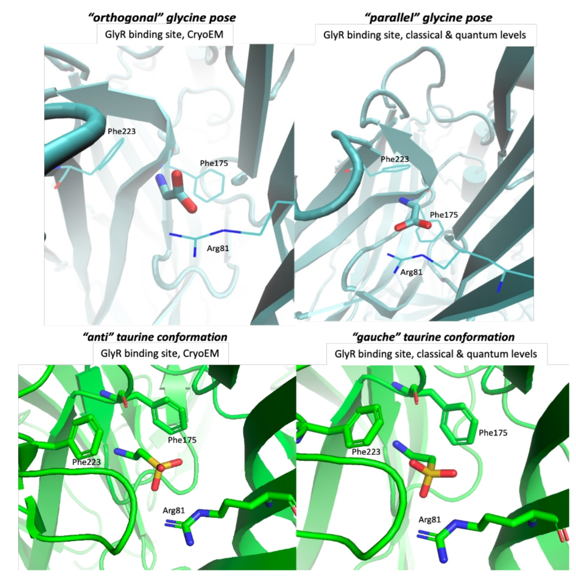
Figure 1: Close-up views of the receptor's docking pocket. Left: the positions of glycine (top) and taurine (bottom) suggested by cryo-electron microscopy images. Right: the positions found by both classical and quantum-mechanical computer simulations. In the simulations, glycine turns so that its oxygen atoms face the receptor's arginine 81 side by side, and taurine twists into a more compact shape. Labels such as Phe175 and Arg81 mark individual building blocks of the receptor. Image: H. Sinenka, D. Mandelli et al., Forschungszentrum Jülich / University of Strasbourg.
Why a supercomputer was needed
The most reliable judge is quantum mechanics, the fundamental theory describing how electrons behave and hold molecules together. Quantum calculations do not depend on simplified, pre-fitted rules, but they are extremely costly. Treating an entire receptor embedded in a cell membrane and surrounded by water at this level is impossible even on today's largest computers. The team therefore used a hybrid strategy: the docking pocket, consisting of the messenger molecule and the twelve surrounding protein building blocks, was described with quantum mechanics, while the rest of the receptor, the membrane and the water were treated with a classical model. Special corrections were added so that the weak but decisive attractions in the pocket were captured accurately.
Even with this shortcut, each simulation required following the motion of every atom in steps of less than a millionth of a billionth of a second, with the electronic structure of the pocket recalculated at every single step. To obtain statistically reliable answers, the researchers ran two dozen independent simulations for four different starting situations, with and without water molecules inside the pocket. This was only possible on the JUWELS CPU Module at the Jülich Supercomputing Centre, using the highly scalable MiMiC software, which lets a quantum program and a classical program run side by side on separate groups of processors. Depending on the system, between 768 and 1,200 processor cores worked together on each calculation.
Results
The verdict of the quantum simulations was clear. In about three out of four cases, taurine adopted the same twisted shape that the classical simulations had predicted, whereas only one of the five experimental structures showed it. Likewise, glycine settled into the same rotated position relative to arginine 81 in about three out of four cases, a position that none of the fourteen experimental structures displayed. In both cases the rearrangement happened within the first trillionths of a second, just as in the classical simulations. Whether or not water molecules were present in the pocket made no significant difference.
These findings show that the disagreement between experiment and simulation was not caused by shortcomings of the classical models, but most likely by the limited sharpness of the experimental images in the docking pocket. This is good news for drug discovery: classical simulations are vastly cheaper than quantum ones and are the workhorse for predicting how strongly a candidate drug binds to its target. The study provides strong evidence that these tools can be trusted for the glycine receptor.
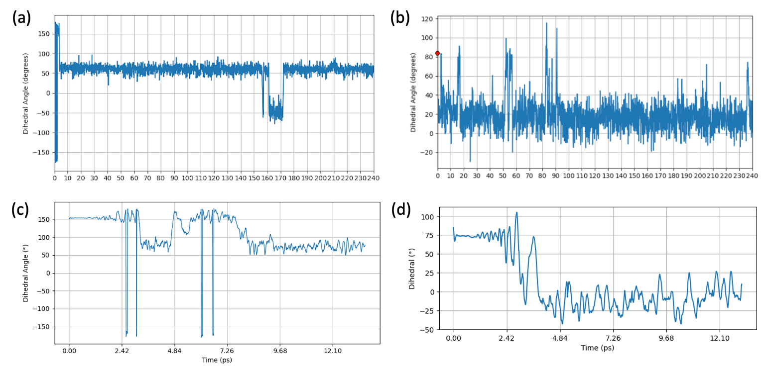
Figure 2: How the messenger molecules rearrange over time. Each curve tracks an angle that describes the shape of taurine (a, c) or the orientation of glycine relative to arginine 81 (b, d) during a simulation; time runs from left to right, in trillionths of a second (picoseconds). Top: classical simulations. Bottom: quantum-mechanical simulations on JUWELS. In both, the molecules leave their experimental starting position within a few picoseconds and settle into a new, stable arrangement. Image: H. Sinenka, D. Mandelli et al., Forschungszentrum Jülich / University of Strasbourg.
Who benefits
The results benefit researchers who design new molecules that act on the glycine receptor, as well as structural biologists who refine experimental models of receptors like it. Knowing the correct position of a molecule in the docking pocket is an essential starting point for developing medicines against hyperekplexia, chronic pain and related neurological conditions. More broadly, the work demonstrates how quantum simulations on supercomputers can serve as an independent referee whenever experiments and everyday simulations disagree.
References
H. Sinenka, “Conception rationnelle des agonistes allostériques des récepteurs synaptiques”, doctoral thesis, University of Strasbourg, defended 17 November 2025. [URL to be added]
Scientific publications based on this project are currently in preparation.
MiMiC QM/MM framework: J. M. H. Olsen et al., J. Chem. Theory Comput. 15, 3810 (2019); V. Bolnykh et al., J. Chem. Theory Comput. 15, 5601 (2019).