
LIFE SCIENCES
Molecular Mechanisms of E-Cadherin Mediated Mechano-Sensing
Principal Investigator:
Frauke Gräter
Affiliation:
Interdisciplinary Center for Scientific Computing, Heidelberg University; and Group for Molecular Biomechanics, Heidelberg Institute for Theoretical Studies
Local Project ID:
chhd33
HPC Platform used:
JUWELS of JSC
Date published:
Feeling the force: molecular mechanisms at cell-cell contacts from simulations
Cells communicate with each other through biochemical as well as mechanical signals. Essential biological processes such as cell division are critically steered by the tension across the cell-cell contacts. We analyzed the underlying molecular principles of mechano-sensing at cell-cell contacts using extensive Molecular Dynamics (MD) simulations performed at the GCS.
To this end, we constructed at atomistic resolution the molecular assembly that propagates the force between cells into the cell, consisting of the major players (proteins E-Cadherin, the membrane, p120 and alpha-catenin). We applied pulling forces by adding virtual springs into the simulation system, mimicking the situation in a stretched cell. The 'softest' building block of the protein assembly, E-cadherin, straightened first, resulting in important changes in interactions of the more 'stiffer' proteins within the assembly. Our simulations can give first insights into how these proteins present at the cell-cell contact change their structure and localization and thereby help to sense mechanical stimuli. Our findings can help understanding the mechanisms by which tissues such as skin grow along the direction of force.

Legend: p120 dynamics under force. While E-cadherin (Transmembrane helix in purple and disordered tail in blue, cyan and yellow) was under force (red arrow), p120 (green) was strongly rotated relative to the membrane, prefers a wide range of relatively flat orientations, which allows membrane interactions and rotationally constraints it to finally 40-50 degrees. On the other hand, this affects interfacial area of the unspecific binding site between E-cadherin (dark blue) and p120, DSASA, shows large fluctuations upon stretching the E-cadherin tail. © UHD and HITS
1 Abstract
E-cadherin provides the mechanical inter-cellular link at adherens junctions and serves as a dynamic hub that translates cell-cell adhesion forces into biochemical downstream processes, the molecular mechanisms of which remain to be explored. The intrinsically disordered cytoplasmic tail of E-cadherin binds to p120, b-catenin, and indirectly to acatenin and actin. Mechanical force leads to a displacement of p120 and b-catenin from the E-cadherin tail, processes which are critical for tension-regulated cell division. The proposed work aims at unravelling the mechanisms underlying these E-cadherin mechano-sensing functions, using atomistic simulations and bioinformatics analyses.
We hypothesize that the E-cadherin tail uncoils and straightens under the tensile force of adherens junctions, and thereby reorients the attached elongated armadillo-repeat catenin molecules along the force direction, which in turn directly affects the E-cadherin/catenin interactions. We will test this hypothesis by constructing atomistically resolved models of the E-cadherin transmembrane and cytosolic domains embedded into the membrane, and successively adding the catenin binding partners to the disordered E-cadherin tail. We will then subject these structures to MD simulations in presence and absence of a tensile forces, mimicking the adherens junction conditions. Given the comparably short Ecadherin cytosolic tail and the rotational constraint the mechanical force has on the bound catenins and their binding interfaces, we expect the force-depending dynamics obtained from simulations to show substantial changes in the cadherin/catenin interactions. Such changes could manifest themselves as an altered p120-membrane interaction, steric hindrance among the two adjacently bound p120 and b-catenin molecules, or a decrease of interfacial interactions at the E-cadherin/catenin binding sites. We will additionally also address the effect of E-cadherin phosphorylation on these processes by additional simulations with modified phosphorylation states. Finally, we will ask whether the observed mechano-sensing mechanisms are similarly at play among the large families of conventional cadherins and catenins, by large-scale sequence alignments and coevolution analysis.
2 Scientific work accomplished and results obtained
In this granting period, we had performed MD simulations by using GROMACS package on:
1. A membrane system with E-cadherin, p120, and tested under force.
2. System 1 but without membrane, and tested under force.
3. System in 1, with b-catenin attached to E-cadherin, and tested under force.
4. A membrane system with E-cadherin, LGN, and tested under force.
Simulations were performed on HPC system JUWELS of JSC.
2.1 Characterizing the cadherin-catenin dynamics
We had performed MD simulations for system 1 (A membrane system with E-cadherin, p120, and tested under force, for 3 times). The major finding in this subproject is the role of membrane and the binding affinity of E-cadherin with p120. We found in pulling simulations (Fig. 1B), p120 will repeatedly interacted/beat the membrane, and resulted the fluctuating of the binding affinity (represented by DSASA) between p120 and E-cadherin.

Fig. 1: p120 dynamics under force. (a) Representative membrane/JMD/p120 structures without (left) and with (right) stretch. (b) Schematic view of P120 orientation with respect to the membrane. The red arrow shows the direction of the pulling force on the E-cadherin tail. Black arrows define the angle shown in b. (c) Orientational angle of P120 relative to the membrane during equilibration (black) and a representative pulling (red) simulations. P120 prefers a wide range of relatively flat orientations, which allows membrane interactions, while force strongly rotates the protein and rotationally constraints it to finally 40-50 degrees. (d) Interfacial area of the unspecific binding site between E-cadherin (dark blue in c) and p120 (green in c), DSASA, shows large fluctuations upon stretching the E-cadherin tail. © UHD and HITS
In this case, we find some limitations of the forcefield: 1. The IDP tail of E-cadherin (Fig. 1A yellow) has a trend to be coiled; 2. P120 tend to bind with membrane. These might be unrealistic. To partially solve this, we performed MD for system without membrane. (System 2)

Fig. 2: p120 (without membrane) dynamics under force. In pulling simulations, the structure of p120 remain stable (A and B). But the interaction area decreases without the fluctuating behavior. (C and D) © UHD and HITS
We have observed optimized accuracy of simulations and almost solve the above-mentioned problems and determine the role of membrane in affinity change of p120 under force. This makes the replacement of p120 by LGN possible. (subproject 2.2) Before we continue with subproject 2.2, we try to test the system 3, which b-catenin included in the system. Our major interests are to test whether there is steric clash in pulling simulations or still the affinity change between different proteins. Our results are consistent with the formal simulations. It is fluctuating of affinity between E-cadherin and p120/bcatenin. (Fig. 3)

Fig. 3: p120 and b-catenin dynamics under force. A-F show behavior of p120/b-catenin with E-cadherin under force. We have not observed steric clash between different players under force, but the affinity changes. © UHD and HITS
2.2 Mechano-sensing mechanism of E-cadherin-LGN binding for force-aligned cell division
By constructing homoly models of LGN with E-cadherin, we investigated the dynamics of LGN under force. From initial results (Fig. 4), we have observed less membrane instractions and the binding affinity between E-cadherin and LGN decrease linearly over time. These results confirmed the bining of LGN is more stable then p120 under force. Next step is to produce/repeat more simulations and analyze the details of simualtions.
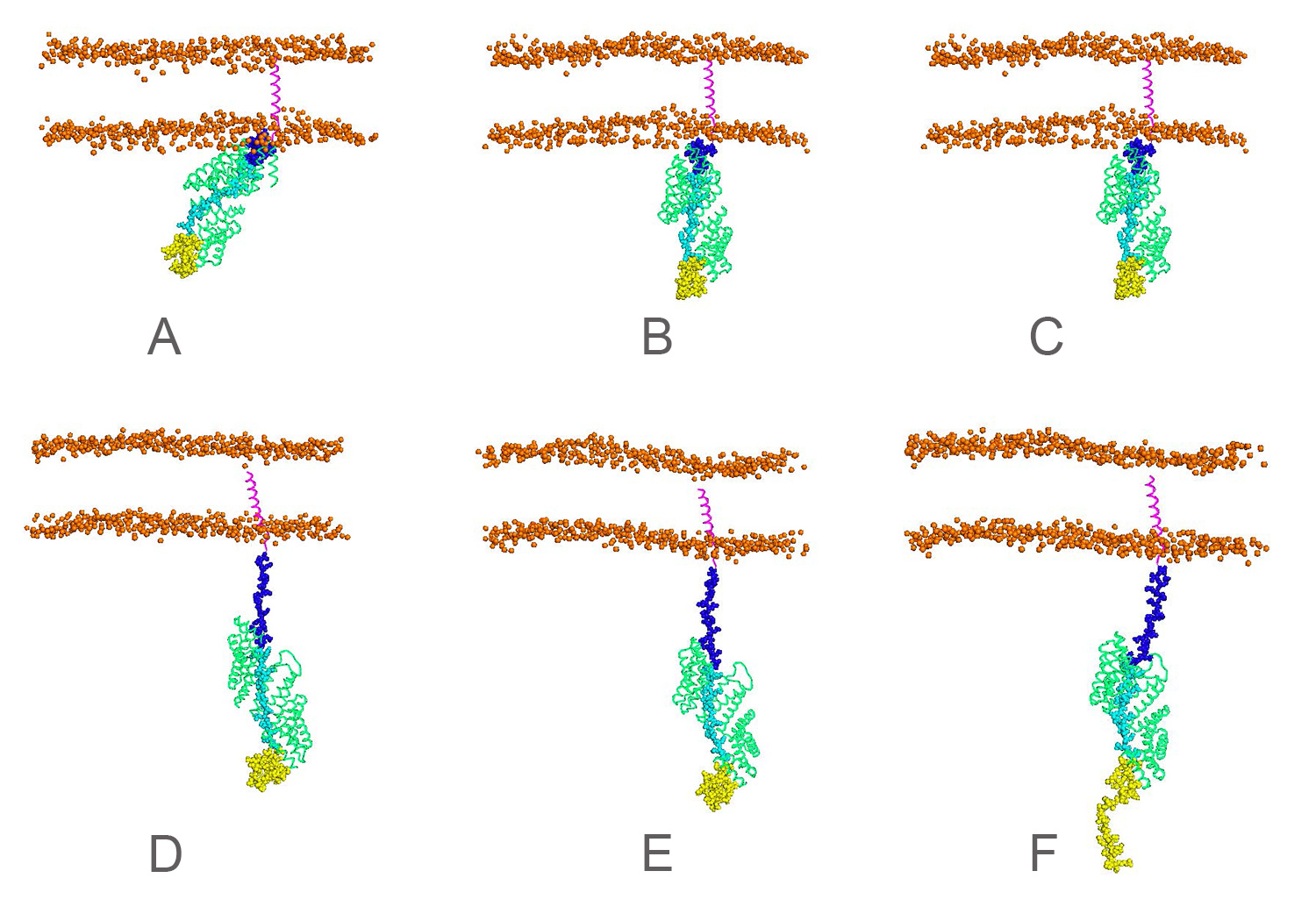
Fig. 4: LGN dynamics under force. In pulling simulations (A to F), the structure of LGN (green) remain stable but show less membrane interactions. © UHD and HITS
3 Realization of the project
We have performed MD simulations by using GROMACS for these systems:
1. A membrane system with E-cadherin, p120, 2 initial structures (equilibrate three initial structures, 256 cores) and tested under force (2 * 3 three velocities 1m/s, 0.1 m/s, 0.033 m/s, 512 cores).
2. System 1 but without membrane, (equilibrate one initial structures, 128 cores) and tested under force (1 * 3 three velocities 1m/s, 0.1 m/s, 0.033 m/s, 256 cores)
3. System in 1, with b-catenin attached to E-cadherin (equilibrate 1 initial structure 512 cores) and tested under force. (1 * 3 three velocities 1m/s, 0.1 m/s, 0.033 m/s, 1024 cores)
4. A membrane system with E-cadherin, LGN, (equilibrate one initial structures, 256 cores) and tested under force (1 * 3 three velocities 1m/s, 0.1 m/s, 0.033 m/s, 512 cores).
This project is rather challenging, because the large membrane/protein system requires lots of computing time. On the other hand, to assemble the membrane-transmembrane+IDP tail-p120 system is not easy: it is a combination of x-ray/NMR structure and MD equilibrated structure. Another difficulty is to find the most possible orientation of the system through MD simulations (conformational sampling).
The most important finding in this project is to combine to “macro” cell experiment with “micro” atomistic MD simulations for E-cadherin - p120/other catenin. Our results provide insight on why E-cadherin bind with LGN stronger than p120 under force, which in turn shed light on the mechanism of E-cadherin mechano-sensing functions and daughter cells appear in the direction of pulling. Thus we believe our project will have a wide readership: people who work on mechano-sensing and developmental biology as well as membrane simulations, and pulling simulations will be interested in our work.
Publications
1. Fan Jin and Frauke Graeter. How multisite phosphorylation impacts the global conformation of IDPs? To be submitted July 2020.
2. Fan Jin and Frauke Graeter. Molecular mechanisms of E-Cadherin mediated mechano-sensing. In preparation.
Research Team
Frauke Graeter (PI) and Fan Jin (both: Interdisciplinary Center for Scientific Computing, Heidelberg University, Group for Molecular Biomechanics and Heidelberg Institute for Theoretical
Studies)
Scientific Contact
Prof. Dr. Frauke Gräter
Interdisciplinary Center for Scientific Computing
Heidelberg University (UHD) - Group for Molecular Biomechanics, and
Heidelberg Institute for Theoretical Studies (HITS)
Schloß-Wolfsbrunnenweg 35, D-69118 Heidelberg (Germany)
e-mail: frauke.graeter [@] h-its.org
LRZ project ID: chhd33
July 2020